#Retina treatment in Mumbai
Explore tagged Tumblr posts
Text

Retina Treatment in Mumbai / Best Retina Treatment in Mumbai
URL- https://www.arohieye.in/eye-care-specialities/retinal-diseases
Your ability to engage with the outside world depends on your eyes. You can see because the components of the eye function together. Many eye illnesses and injuries can affect how the eyes function. Arohi Eye Hospital is one of the most renowned that has been helping innumerable patients cure various types of eye diseases for the past few years. If you’re suffering from any retina-related problem, it is crucial to get a retina treatment in Mumbai right away as it calls for a medical emergency
About Arohi Eye Hospital The high-end technology, and the dedication and experience of the doctors and staff has proven Arohi Eye Hospital to be the best eye hospital in Mumbai, having served more than 50,000 patients for an array of different eye diseases and disorders, each having successfully accomplished the results required.
0 notes
Text
Retina Treatment in Mumbai
Situated in Mumbai, the "Raghunath Netralaya" is an ultra-specialist retina care facility "exclusively" dedicated to treating conditions affecting the posterior segment of the retina. Our organisation specialises in treating medical and surgical retinal problems, ocular imaging techniques, and uveitis, also referred to as ocular inflammation.
#Retina Treatment Mumbai#Best Retina Specialist Doctor Mumbai#Cornea Treatment Mumbai#Cornea Transplant Surgeons Mumbai#Cataract Treatment Mumbai#Cataract Operation Mumbai#Eye Care Hospital Mumbai#Best Eye Doctor Mumbai#Top Eye Hospital Mumbai
0 notes
Text
Comprehensive Retina Care at Shreeji Eye Clinic
Finding the right care for your retina is essential for preserving your vision. At Shreeji Eye Clinic, our team of the best retina specialists in Mumbai ensures that each patient receives personalized care and attention. With years of experience and a patient-centric approach, our specialists are trusted by countless individuals across Mumbai and beyond.
#Best Retina Specialist in Mumbai#Retina Laser and Surgery Mumbai#Best Retina Surgeons in Mumbai#Retina Laser Surgery Doctors In Mumbai#Mumbai Eye Retina Clinic#Best Retina Treatment In Mumbai#keratoconus treatment mira road#Keratoconus Treatment in Mumbai#best retina specialists in Mumbai#retina laser and surgery in Mumbai
0 notes
Text
Understanding Diabetic Retinopathy
Diabetic retinopathy (DR) sits at the top of the main causes found for vision loss in people with diabetes. It occurs when high blood sugar levels damage the delicate blood vessels in the retina. This damage can lead to bleeding, swelling, and even the growth of abnormal new blood vessels, all of which can significantly impair your vision.
The good news? Diabetic retinopathy is largely preventable with proper diabetes management and early detection. Millions of people worldwide are affected by DR, but with regular eye exams and timely treatment, the risk of vision loss can be dramatically reduced. Continue reading this blog to gain an overall understanding around this phenomenon and educate yourself about the best treatment for diabetic retinopathy.
How Does Diabetes Affect Your Eyes?
When blood sugar levels remain high for extended periods, it can wreak havoc on the tiny blood vessels throughout your body, including those in the retina. These delicate vessels become weakened and fragile, leading to a cascade of problems. They may leak fluid or blood into the retinal tissue, causing blurry vision or even complete vision loss in some cases.

Understanding the Different Stages of DR
Diabetic retinopathy progresses through different stages, each with its own set of characteristics:
Nonproliferative Diabetic Retinopathy (NPDR): This is the earlier stage of DR, where the blood vessels in the retina are damaged but vision loss is usually not a concern. However, symptoms like microaneurysms (tiny bulges in the blood vessels), retinal hemorrhages (bleeding), and cotton wool spots (waxy yellow deposits) might be present. Early detection and management of NPDR are crucial to prevent progression to the more advanced stage.
Proliferative Diabetic Retinopathy (PDR): In this advanced stage, the body attempts to compensate for the damaged blood vessels by growing new, abnormal blood vessels on the surface of the retina. These new replacements are fragile and prone to bleeding, which can lead to severe vision loss or even complete blindness if left untreated.
Who's Most at Risk for Diabetic Retinopathy?
Anyone with diabetes, including type 1, type 2, or gestational diabetes (develops during pregnancy), can develop diabetic retinopathy. The longer you have diabetes, the higher your risk. Even factors like poor blood sugar control, high blood pressure and cholesterol, smoking can all further increase your risk of developing DR.
Don't Ignore These Warning Signs: Act on Vision Changes
Diabetic retinopathy can often progress silently, with no noticeable symptoms in the early stages. However, as the condition worsens, you may experience changes in your vision, such as:
Blurred or distorted vision
Seeing dark spots or floaters in your field of vision
Sudden vision loss, especially in one eye
Difficulty seeing at night or in low light conditions
Eye pain or redness
If you experience any of the symptoms mentioned above, please see a diabetic retina specialist as soon as you can.
Taking Control: Treatment Options for DR
Fortunately, there are effective treatment options available for diabetic retinopathy. The condition specific treatment you receive will depend on its severity. Here are some common methods used by diabetic retinopathy specialists:
Medications: Anti-VEGF medications can be injected into the eye to help reduce swelling and prevent blood vessel leakage. Corticosteroid injections might also be used to manage inflammation.
Laser Treatment: Laser therapy can be used to seal leaking blood vessels or destroy abnormal new blood vessels to prevent bleeding and further damage to the retina.
Vitrectomy: In some advanced cases, surgery called a vitrectomy may be necessary. This procedure involves removing blood or scar tissue from the vitreous gel, the clear jelly-like substance that fills the center of your eye.
Living with Diabetes and Protecting Your Sight
Diabetic retinopathy can be a scary diagnosis, but with proper diabetes management, regular eye exams, and timely treatment, you can significantly reduce your risk of vision loss. Millions of people worldwide manage diabetes and DR successfully. Therefore, to simplify the process of finding the best eye doctor in Mumbai for diabetic retinopathy, you can simply contact Shri Venkatesh Eye Institute. Their active role in your healthcare and important recommendations will help protect your vision, allowing you to enjoy a safer future.
#best eye surgeon in mumbai#best treatment for diabetic retinopathy#best eye doctor for diabetic retinopathy#diabetic retina specialist#diabetic retinopathy specialists#best doctor for diabetic retinopathy
0 notes
Text
What are the tests Performed to Diagnose Glaucoma?

Several tests are performed to diagnose glaucoma, a group of eye conditions that can lead to optic nerve damage and vision loss. The specific tests may vary based on the suspected type of glaucoma and the individual's eye health. Here are common tests performed to diagnose glaucoma:
1-Tonometry:
Measures intraocular pressure (IOP), which is a key risk factor for glaucoma. Elevated IOP can indicate a higher risk of glaucoma, although normal IOP does not rule out the condition.
2-Ophthalmoscopy:
Allows the eye care professional to examine the optic nerve for signs of damage. Changes in the appearance of the optic nerve head, such as cupping, may suggest glaucoma.
3-Visual Field Test (Perimetry):
Assesses the full horizontal and vertical range of what a person can see in their field of vision. Glaucoma often leads to peripheral vision loss, which can be detected through this test.
4-Gonioscopy:
Examines the drainage angle of the eye to determine if it is open or closed. This helps in classifying the type of glaucoma, such as open-angle or angle-closure glaucoma.
5-Pachymetry:
Measures the thickness of the cornea. Corneal thickness can influence intraocular pressure readings, and thin corneas may be associated with a higher risk of glaucoma.
6-Optical Coherence Tomography (OCT):
Uses light waves to create detailed cross-sectional images of the optic nerve and retina. It helps in assessing the thickness of the retinal nerve fiber layer, which can be indicative of glaucomatous damage.
7-Retinal Imaging:
Captures high-resolution images of the retina, helping to detect any structural changes or damage related to glaucoma.
8-Visual Acuity Test:
Measures how well an individual can see at various distances using an eye chart. While not specific to glaucoma, changes in visual acuity may be observed in advanced stages of the condition.
9-Corneal Hysteresis Measurement:
Evaluates the cornea's ability to absorb and return energy during deformation. This measurement can provide additional information about the biomechanical properties of the cornea and the risk of glaucoma progression.
It's important to note that glaucoma diagnosis is often a combination of these tests, and regular eye examinations are crucial, especially for individuals at higher risk, such as those with a family history of glaucoma or individuals over the age of 40. If you have concerns about your eye health, consult with an eye care professional for a comprehensive examination and appropriate testing.
For more information, Consult Dr. Vaidya Eye Centre as they provide best Glaucoma Treatment in Mumbai or you can contact us on 9004496621.
#dr. vaidya eye centre#drvaidyaeyehospital#eye specialist in andheri#eyespecialist#eye clinic in andheri#eye hospital in mumbai#eyecare#eyehealth#glaucoma treatment in mumbai#retina specialist in mumbai#Glaucoma Treatment#Glaucoma#Eye Centre#glaucoma diagnosis
0 notes
Text
Retinopathy of prematurity (ROP) - Ojaseyehospital
Retinopathy of prematurity (ROP) is a potentially blinding disease caused by abnormal development of retinal blood vessels in premature infants. Infants weighing about 2¾ pounds (1250 grams) or less and are born before 31 weeks of gestation are more likely to be affected by ROP. Retina of the eye is the innermost layer, that receives light and turns it into visual information that is sent to the brain.
In Premature babies, the blood vessels that feed the retina haven’t finished growing, and get disturbed growth pattern due to premature birth. When ROP is severe, it can cause retinal detachment and possible blindness. Smaller a baby at birth, more likely chances are that baby will develop ROP. This disorder affects mostly both eyes and is the most common cause of vision loss in childhood and can lead to lifelong vision impairment and blindness.
How common is ROP?
With recent advances in neonatal care, premature babies are saved and not all premature infants develop ROP. Out of 20 million children born annually in India and 0.6 million admitted to NICUs all over the country, about 10% of these infants are at risk of developing ROP. As per the figure given by Clare Gilbert in 2010, there are about 0.3 million blind children (>15 years of age) in India and 10% of them are blind due to ROP. There are approximately 3.9 million infants born in the U.S. each year. About 14,000 are affected by ROP and 400-600 infants each year in the U.S. become legally blind from ROP. About 90 percent of all infants with ROP are in the milder category and do not need treatment.
How ROP Develops?
Eye formation starts at 16th week of pregnancy. Blood vessels begin forming around optic nerve. These blood vessels grow towards the edges of the developing retina supplying oxygen and nutrients. In the last 12 weeks, eye matures rapidly and if a baby takes birth prematurely, normal vessel growth may stop and retinal edges may be devoid of nutrients and oxygen. According to some studies, periphery of the retina sends out signals for nourishment and as a result, new abnormal vessels begin to grow. These newly formed vessels are weak and tend to bleed, leading to scarring of retina and ultimately detachment. This is the prime cause for blindness in ROP.
Risk Factors for ROP
Factors contributing to the risk of ROP include anemia, blood transfusions, respiratory distress, breathing difficulties, Hyperoxia, Hypoxia, Hypotension and the overall health of the infant. The most important risk factors for ROP are prematurity and low birth weight. Phototherapy is also attributed to the development of ROP.
Signs of Retinopathy of prematurity:
ROP shows variety of presentations:
Avascular retina
Scarring or dragging of the retina
Vitreous hemorrhage
Cataract Development
Retinal detachment.
Do everyone need to see retina specialist
Premature babies before 30 weeks of gestation and babies who are underweight are at higher risk. These infants are always advised to visit eye doctor for checkup. Best time to visit retina specialist is preferred within 30 days of birth. Retinal screening done in this period is quite beneficial for the treatment of the infant.
Treatment
Mild cases do not need any treatment, while in advanced cases a variety of treatment options are available. The most effective and proven treatment for ROP is laser therapy or cryotherapy.
Late stages may require scleral buckle or Vitrectomy
Scleral buckle – It is usually done in stage IV or V. A silicone band is placed around the eye, It keeps the vitreous gel from pulling on the scar tissue and allows the retina to flatten back down onto the wall of the eye. This band is removed later as eye continues to grow; otherwise infant will develop myopia.
Vitrectomy – This is performed in stage V only and involves removing the vitreous and replacing it with a saline solution. Once vitreous has been removed, the scar tissue on the retina can be peeled back, allowing the retina to relax and lay back down against the eye wall.
Tag = Eye Hospital in Mumbai, Retina treatment in India, best Retina Hospital In Mumbai, Best Eye Surgeon in Mumbai
#Eye Hospital in Mumbai#Retina treatment in India#best Retina Hospital In Mumbai#Best Eye Surgeon in Mumbai
0 notes
Text
Is Diabetic Retinopathy painful?
Diabetic Retinopathy is usually painless and sometimes doesn’t even cause any symptoms in the early stage.
Some symptoms of Diabetic Retinopathy are:
Blurry Vision Loss in Vision Spots in Vision Floaters (dark floating string in vision) Dark areas or empty areas in vision

One of the most effective ways of treating Diabetes and its complications is Sanjeevan Netralaya’s tailor made Ayurvedic treatments that are unique for every patient.
Sanjeevan Netralaya’s treatment has helped lakhs of people across India without adding to the discomfort and pain or any unnatural side effects.
#sanjevan netralaya#eye care#blue eyes#eyesight#eye health#diabetic retinopathy treatment#eye diseases#diabetic retinopathy#retina care center#retina centers#mumbai#opthalmic diseases#india
1 note
·
View note
Link
#Dr. Jatin Ashar#Mumbai Eye Care#Eye Specialist in Ghatkopar#eye care ghatkopar east#cornea specialist in Mumbai#Keratoconus Treatment In Ghatkopar#Retina Specialist in Mumbai
0 notes
Text
7 Best Lasik Eye Surgery Centers in India

Lasik eye surgery has made eyeglasses passé. For who would not want to flaunt their eyes out in the open? I am sure all of us would like to get rid of the cage our eyes have to be continuously in, no matter how stylishly we wear them, we still aspire to be free from them. Lasik eye surgery has given hope to millions who suffer from myopia (nearsightedness), astigmatism (refractive error of the eye), &hypermetropia (farsightedness), and even presbyopia (insufficiency of eyes due to aging) in some cases.
However, any correction or surgery if not required, should not be undertaken, so goes the adage. Like all other surgeries, even this one has its limitations and involves risks. Dry eyes, halos, ocular neuropathic pain, and subconjunctival hemorrhage, are some of the non-serious adverse effects of Lasik surgery.
However, flapped discs, higher-order aberrations, uveitis, slipped flap, retinal detachment, Choroidal neovascularization (CNV) and tendencies of depression and suicide (less commonly observed) in patients can be seen as more severe expressions of risks from the surgery. Thus, "if not required, do not attempt it" is the apparent suggestion good hospitals and doctors would provide you. However, if it becomes mandatory at some point in your life, do make sure you choose from some excellent hospitals where doctors can prevent corneal thinning in the best possible way during the surgery.
Generally, 25-40 years is the apt age to undergo Lasik eye surgery even though the Food and Drugs Administration (FDA) has approved Lasik eye surgery for patients 18 years of age and above in the United States. Doctors generally do not suggest it below 25 years of age except in special cases. Vision correction also does not mean that you will never need glasses or lenses again. This is a myth and instead what is more appropriate to say in this regard, is that your numbers going forth might not be so high as they used to be before.
If you are pregnant or already have thin cornea or a lot of vision change prescriptions, do not attempt Lasik eye surgery. Once your power stabilizes and you do not have any power changes for six months or above, only then try Lasik eye surgery after thorough consultation with your ophthalmologist.
So, without any further delay, let us find out which hospitals can tackle the surgery well in our country. Below we have listed the seven best hospitals for Lasik eye surgery in India.
Foremost Hospitals for Lasik Eye Surgery
Some of the best hospitals where the care provided to patients during/after surgery is worth applauding are being listed below.
1. Sankara Nethralaya
If you wish to avail the latest LASIK eye surgery in India, then you could opt for Sankara Nethralaya. You can have a corneal assessment, expert counseling, pre-and post-operative customized care and even prompt follow-up care.
Types of laser vision correction surgeries available at Sankara Nethralaya comprise of LASIK (Laser In-Situ Keratomileusis), Festo LASIK, PRK (Photorefractive Keratectomy), Epi LASIK (Epithelial Laser-Assisted In-Situ Keratomileusis), and Wave front-guided LASIK.
2. Retina Foundation and Eye Research Center
Retina Foundation is a well-reputed center for undergoing a LASIK eye surgery. They are providing eye care facilities since 1976 and is a pioneer in Vitreo Retinal diseases.
Equipped with modern facilities and an expert panel of ophthalmologists and eye surgeons, the Retina Foundation is the best resort for any eye complains as they have a wide range of treatment and care facilities available.
3. Asian Eye Institute & Laser Centre – Mumbai
Asian Eye Institute & Laser Centre offers an excellent opportunity to undergo LASIK eye surgery and treatment in Mumbai under the guidance of Dry Hitesh Chedi.
Patients with myopia, hyperopia, and astigmatism can get their vision corrected at this facility to a great degree of precision. BASIC, Customized (Wave front guided), and BLADE LESS LASIK surgeries are conducted at this facility.
4. Acuvision Eye Hospital, Eye Clinic in Khari
Acuvision is another reliable and reputed center for LASIK eye surgery and other comprehensive eye care solutions. Advanced technologies, digitalized machines, and a team of expert ophthalmologists help rectify a lot of eye problems.
5. Artemis Hospital, Gurgaon
This multi-speciality hospital also has an ophthalmology center where patients suffering from myopia, hypermetropia, or astigmatism can undergo laser vision correction.
Besides this, Artemis Hospital's ophthalmology department also offers other solutions to rectify power and vision-related problems in patients such as Phaco with Multifocal IOL surgery and Implantable Collamer Lens (ICL) surgery.
6. Bombay City Eye Institute & Research Centre
A center with complete eye care facilities, Bombay City Eye Institute & Research Centre, is another trusted place to get your vision rectified with modern solutions and surgical treatments.
Diagnostic machines like Oculyzer II (known as Pentacam- HR) and Topolyzer Vario are used for analyzing the eye. The department performs refractive corrections and surgeries like bladeless LASIK, PRK (photorefractive eye surgery), ICL (implantable contact lens), CLE (Clear lens extraction) with Trifocal IOL (Trifocal intraocular lenses), and even facilitates advanced Contoura Vision (USFDA (United States Food and Drugs Administration) approved eye laser for specs removal).
7. Maxivision Super Speciality Eye Hospital
With 25 years of experience, Maxvision Eye Hospital offers a range of fixes for refractive eye errors (myopia, hyperopia, astigmatism) such as LASIK/PRK (laser-assisted in situ keratomileusis /Photorefractive keratectomy), Custom LASIK, ICL (implantable Collamer lens), SMILE (Small Incision Lenticule Extraction), and Presbyopic Laser treatments.
So, now that you are aware of which hospitals can help you the best possible way, choose your hospital wisely. Lasik eye surgery should not be attempted as soon as you get your lenses.
If there is something else that you would want to add, please drop your comments in the box below.
2 notes
·
View notes
Text

best cataract surgery in mumbai
What is Cataract?
The lens of the eye which is made of water and protein helps focus light into the retina through which visual signals are sent to the brain. Excess of protein building leads to lens blocking. The cloud form on your eye lens due to excess protein reduces vision due to blockage of light passing to the retina. This excess protein built-up which leads to lens blockage is called cataract.
Who can have Cataract?
Cataract is considered as a normal process of aging. At our center for cataract treatment we come across patients from the age group of 60 plus. Few patient cases in the age group of 45-60 are also detected for femtosecond Laser Cataract Surgery in Mumbai at our center.
Symptoms of Cataract
Some of the more common symptoms of cataract include:
Glare
Fading or yellowing of colours
Poor night vision
Double vision in one eye
Halos around lights
A feeling of viewing through a frosted piece of glass or fog
Blurred vision
Frequent change in eye glasses or contact lenses
If you are facing such symptoms then an early detection of cataract will help you take a decision under the professional guidance of our Doctors.
People with a cataract in only one eye may notice a loss of depth perception; this can cause problems in judging where stairs are and determining the distance of cars driving in front of them.

lens replacement surgery render accurate correction for vision. If you would like to be femtosecond laser and it will also a leading reason why cataract surgeons are excited about their potential, due to the automation these lasers can provide – in creating the capsulorhexis and in pre-chopping the nucleus, reducing the overall energy needed to remove the cataract.
Why The Vission Eye Center for Cataract Surgery Treatment?
Our surgeons are all highly trained and have had extensive experience with the most up-to-date techniques used for cataract surgery in Mumbai at our center. The OSC offers a relaxed and friendly setting for the patient and the patient’s family. Shortly after cataract operation is completed, the patient may go home and resume almost all routine activities.
It must be understood that complications may occur in all types of surgery. In cataract treatment, haemorrhage, infection, and swelling are all possible, but very uncommon. The chance of any significant cataract surgery complications is less than 1%. It is among the safest and most successful procedure in the medical field. However, if a problem does arise, prompt treatment may resolve it.
About a year after surgery, approximately 20% of the patients who undergo femtosecond Laser cataract surgery develop a haze of the capsular membrane surrounding the lens implant. Should this occur, YAG laser treatment is recommended. The YAG laser is used to create an opening in the clouded membrane, which significantly improves the patient’s vision. It is one of the safest treatments used in ophthalmology. It is painless, requires no anaesthesia or incision, and takes only minutes to complete.
Laser in cataract render accurate correction for vision. Laser in cataract also is a leading reason why the best cataract surgeons in Mumbai are excited about their potential. Due to the automation, these lasers can provide – in creating the capsulorhexis and in pre-chopping the nucleus, reducing the overall energy needed to remove the cataract. Depending on the growth of cataract & the damage it causes to your vision, the call to remove it is taken. Our consultation process gives you an opportunity to understand cataract surgery or procedure involved, the cataract operation cost in Mumbai. A larger portion of cloudiness in your eye lens can impair vision partially or completely. In such cases, it is advisable to have a cornea transplant in Mumbai at our clinic, The Vission Eye Center.
Cataract Surgery Treatment
Presently, there is no medication, eye drops, exercises or glasses to cure or prevent cataracts. Surgery is the only way to remove a cataract. Cataract surgery is one of the most safest and common type of surgery. Cataracts cannot be removed with a laser, only through surgical incision. In cataract surgery the cloudy lens is removed from the eye.
The focusing power of the removed lens is achieved by replacing it with a permanent intraocular lens implant (IOL), which has been selected to suit the specific eye measurements of each patient. The expense of Cataract surgery is an essential subject on the grounds that it is the most regularly performed surgery on the planet.
0 notes
Text
Arohi Eye Hospital is one of the best for Retina Treatment in Mumbai
The Hospital is well known for its top retina specialists that are highly trained in diagnosing and treating retina conditions such as diabetic retinopathy, age-related macular degeneration (AMD), and retinal detachment, which can cause vision loss or blindness. So, if you're experiencing any issues with your retina, our retina eye specialist are always there to assist you and ensure that you get the best possible eye care.
Common Retinal Diseases- Floaters, Diabetic Eye Disease, Retinal Detatchment
0 notes
Text
Retina Specialist
Retina Specialist in Andheri , Mumbai
UNDERSTANDING RETINAL DISEASES
Home
Treatments
Retina Specialist
Retina Specialist in Mumbai
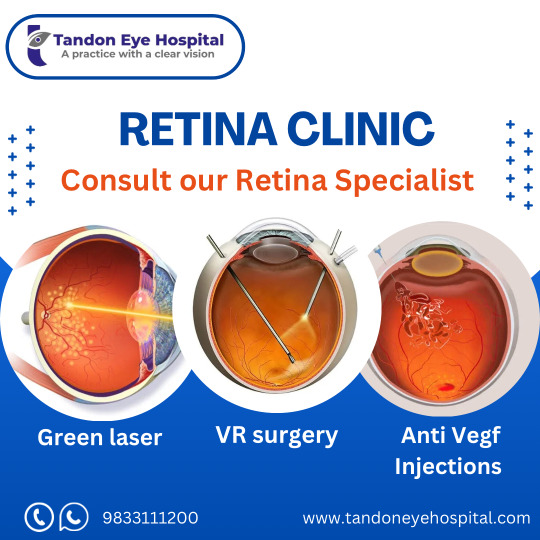
At Tandon Eye Hospital, we realize how retinal diseases can impact your sight and life in general. The retina is a thin layer of tissue at the back of the eye that critically converts light into visual signals for the brain. Damage to the critical tissue here will surely cause severe loss of vision or complete blindness. Our diagnosis and specialized treatment capabilities treat a wide spectrum of retinal conditions, so you can be assured that you are receiving the best possible care from the best Retina Specialist in Mumbai.

Understanding Retinal Diseases
Retinal diseases can come in many different forms, which uniquely will affect your vision. Some common retinal diseases are:
Retinal Tear: A retinal tear results from the shrinking of vitreous gel within the eye that pulls on the retina, leaving breaks in the tissue. Symptoms usually appear quickly, with sudden floaters and flashes of light.

Retinal Detachment: This is a serious condition in which fluid gathers beneath the retina, lifting it off its normal location. Symptoms can be sudden changes in vision and increased floater.


Vision after retinal detachment often appears blurred or distorted, with potential dark spots, flashes of light, or a curtain-like shadow blocking part of the visual field.
Diabetic Retinopathy: Diabetes can damage the smallest blood vessels in the retina, sometimes causing swelling, bleeding, or even significant vision loss.

Macular Degeneration: This is a progressive disease that affects the central part of the retina, with blurred or distorted vision. It typically affects elderly adults and can either be dry macular degeneration or wet macular degeneration.

Symptoms to Watch For
There are a few symptoms of retinal disease, which you should be aware of:
Seeing floaters or spots in your field of vision
Blurred or distorted vision
Loss of central or peripheral vision
Flashes of light or sudden vision change
If you experience any of these symptoms, consult a Retina specialist as soon as possible.

Diagnostic Procedures
At Tandon Eye Hospital, our expert ophthalmologists, and Retina Specalist, use the newest diagnostic technology to check up on your retinal health. The following diagnostic tests are most important.
Amsler Grid Test: A quick screening tool intended to assess the clarity of central vision and detect distortion.
Optical Coherence Tomography (OCT): This imaging modality captures high-resolution images of the retina for the diagnosis and monitoring of retinal diseases.
Fluorescein Angiography: This imaging modality captures high-resolution images of the retina for the diagnosis and monitoring of retinal diseases.
Indocyanine Green Angiography: It uses infrared light to have a deeper view of the retinal blood vessels.

Treatment Options
Our overall aim is to prevent or slow the progression of retinal diseases and preserve your vision. Treatment options available at Tandon Eye Hospital include:
Laser Surgery: Effective for repairing retinal tears or holes, preventing further detachment.
Cryopexy: A freezing treatment used to secure the retina in place.
Scleral Buckling: A form of surgery in which a silicone band around the eye relieves pressure on the retina.
Vitrectomy: It is a procedure needed to remove the vitreous gel. This is typically performed for severe retinal detachment or with active and uncontrollable hemorrhage.
Intravitreal Injections: Medications are directly injected into the eye, which may be used in the treatment of certain diseases, like age-related macular degeneration, among others.

0 notes
Text
Retina Treatment in Mumbai
Situated in Mumbai, the "Raghunath Netralaya" is an ultra-specialist retina care facility "exclusively" dedicated to treating conditions affecting the posterior segment of the retina. Our organisation specialises in treating medical and surgical retinal problems, ocular imaging techniques, and uveitis,also referred to as ocular inflammation.
#Retina Treatment Mumbai#Best Retina Specialist Doctor Mumbai#Cornea Treatment Mumbai#Cornea Transplant Surgeons Mumbai#Cataract Treatment Mumbai#Cataract Operation Mumbai#Eye Care Hospital Mumbai#Best Eye Doctor Mumbai#Top Eye Hospital Mumbai
0 notes
Text
Achieve Perfect Vision: LASIK Surgery in Mumbai at Krishna Eye Centre

In a world where clear vision is essential, LASIK surgery has emerged as a life-changing solution for those seeking freedom from glasses or contact lenses. At Krishna Eye Centre, recognized as the best eye hospital in Mumbai, we offer advanced LASIK surgery to help you achieve sharp, natural vision with ease.
What Is LASIK Surgery?
LASIK (Laser-Assisted In Situ Keratomileusis) is a cutting-edge refractive procedure designed to correct common vision problems such as myopia (nearsightedness), hyperopia (farsightedness), and astigmatism. The surgery uses precision lasers to reshape the cornea, allowing light to focus correctly on the retina, resulting in improved clarity of vision.
Why Choose Krishna Eye Centre for LASIK Surgery in Mumbai?
When it comes to your eyes, expertise and technology matter. Krishna Eye Centre has established itself as a trusted destination for LASIK surgery in Mumbai, combining skilled professionals with state-of-the-art facilities.
Here’s why Krishna Eye Centre stands out:
Experienced Surgeons: Our team comprises highly qualified ophthalmologists with extensive experience in performing LASIK and other refractive surgeries.
Advanced Technology: We use the latest laser technology to ensure precision, safety, and effective outcomes.
Personalized Care: Every patient’s eyes are unique. We conduct thorough evaluations to design a customized treatment plan tailored to your needs.
Comprehensive Follow-Up: Post-surgery care is as important as the procedure itself. We ensure your recovery is smooth and your vision remains optimal.
Proven Track Record: Thousands of satisfied patients have trusted us for their LASIK surgery in Mumbai, reaffirming our position as the best eye hospital in Mumbai.
What to Expect During LASIK Surgery
The LASIK journey at Krishna Eye Centre begins with an in-depth consultation. Our specialists will perform a series of tests to determine your eligibility for the procedure.
On the day of surgery, here’s what you can expect:
Preparation: Your eye will be numbed with anesthetic drops to ensure comfort.
Procedure: A specialized laser will create a thin corneal flap, which is lifted to reshape the underlying cornea with precision laser technology. The flap is then repositioned.
Quick Recovery: The procedure typically takes 15 minutes for both eyes, and most patients notice significant vision improvement within 24 hours.
Benefits of LASIK Surgery
Immediate improvement in vision
Painless and quick procedure
Long-lasting results
Freedom from glasses and contact lenses
Is LASIK Right for You?
LASIK is a safe and effective option for most individuals, but certain factors such as age, corneal thickness, and overall eye health play a role in determining eligibility. A consultation at Krishna Eye Centre will provide clarity and ensure you’re making the best choice for your vision.
Book Your Consultation Today
If you’re tired of relying on glasses or contact lenses, it’s time to explore LASIK surgery at Krishna Eye Centre. As the best eye hospital in Mumbai, we’re committed to delivering exceptional care and life-changing results. Take the first step towards a clearer future by scheduling your consultation today.
#eye surgery#eye treatment#eye hospital#hospital#LASIKSurgery#MumbaiEyeCare#KrishnaEyeCentre#ClearVision#EyeHealth
0 notes
Text
Understanding Blurry Vision in One Eye: Causes and Treatments Blurry vision in one eye can disrupt daily life, from reading to driving. This guide delves into common and serious causes, including dry eyes, retinal detachment, and glaucoma. Learn about treatments, prevention tips, and the importance of early diagnosis. Discover specialised eye care services like cataract, glaucoma, and retina treatment in Mumbai for better vision. Read more!
0 notes